


Approach ConsiderationsMedical care for heart failure (HF) includes a number of nonpharmacologic, pharmacologic, and invasive strategies to limit and reverse its manifestations. [3, 8, 116] Depending on the severity of the illness, nonpharmacologic therapies include dietary sodium and fluid restriction; physical activity as appropriate; and attention to weight gain. Pharmacologic therapies include the use of diuretics, vasodilators, inotropic agents, anticoagulants, beta blockers, angiotensin-converting enzyme inhibitors (ACEIs), angiotensin receptor blockers (ARBs), calcium channel blockers (CCBs), digoxin, nitrates, B-type natriuretic peptides (BNPs), I(f) inhibitors, angiotensin receptor-neprilysin inhibitors (ARNIs), soluble guanylate cyclase stimulators, sodium-glucose cotransporter-2 inhibitors (SGLT2Is), and mineralocorticoid receptor antagonists (MRAs). Show
2022 ACC/AHA/HFSA GuidelinesUpdated and revised guidelines on the management of HF were published in May 2022 by the American College of Cardiology, American Heart Association, and Heart Failure Society of America (ACC/AHA/HFSA). [4, 5, 6, 7] These guidelines replace the 2013 and 2017 recommendations with significant and paradigm-shifting additional treatment options that include new/repurposed drug therapies that benefit almost without regard to ejection fraction (EF); additional disease-staging terminology that characterizes HF as a continuum; updated recommendations for advanced HF, acute HF, and comorbidities; as well as other guidance. Top 10 key points
2021 ESC GuidelinesClinical practice guidelines on the management of heart failure were also published by the European Society of Cardiology (ESC) in August 2021. Recommendations include the following [68] :
Invasive therapies for HF include electrophysiologic intervention such as cardiac resynchronization therapy (CRT), pacemakers, and implantable cardioverter-defibrillators (ICDs); revascularization procedures such as coronary artery bypass grafting (CABG) and percutaneous coronary intervention (PCI); valve replacement or repair; and ventricular restoration. [3, 8, 61, 63, 116] Heart transplantation has been the criterion standard for therapy when progressive end-stage heart failure occurs despite maximal medical therapy, when the prognosis is poor, and when there is no viable therapeutic alternative. [3, 8, 9] However, mechanical circulatory devices such as ventricular assist devices (VADs) and total artificial hearts (TAHs) can bridge the patient to transplantation; in addition, VADs are increasingly being used as permanent therapy. [8] Comorbidities to considerCoronary artery disease Patients with heart failure should be evaluated for coronary artery disease, which can lead to heart failure (see Etiology). Not only may this condition be the underlying cause in up to two thirds of heart failure patients with low ejection fraction, but coronary artery disease may also play a role in the progression of heart failure through mechanisms such as endothelial dysfunction, ischemia, and infarction, among others. [3] Patients with coronary artery disease with modestly reduced ejection fraction and angina have demonstrated symptomatic and survival improvement with CABG in studies; however, the trials did not include individuals with heart failure or those with severely reduced ejection fractions. [3] In patients with angina and ventricular dysfunction, evaluation with coronary angiography should not be delayed (see Catheterization and Angiography). Noninvasive cardiac testing is not recommended in patients with significant ischemic chest pain, as revascularization is advised in these patients independent of their degree of ischemia/viability. [3] Although there are no reports of controlled trials evaluating heart failure without angina and their outcomes with coronary revascularization, surgical revascularization is recommended in those with significant left main stenosis and in those with extensive noninfarcted but hypoperfused and hypocontractile myocardium on noninvasive testing. [3] In patients with heart failure and reduced LVEF but without angina, it has not yet been determined whether routine evaluation of possible myocardial ischemia/viability and coronary artery disease should be performed. [3] For patients with heart failure from LV dysfunction without chest pain and without a history of coronary artery disease, coronary angiography may be useful in young patients to exclude congenital coronary anomalies. However, because clinical outcomes have not been shown to improve in patients without angina, coronary angiography may not be as useful in older patients for evaluating the presence of coronary artery disease. [3] Some experts nonetheless suggest excluding coronary artery disease whenever possible, particularly in the presence of diabetes or other conditions associated with silent myocardial ischemia, because LV function may show improvement with revascularization. [3] In general, if coronary artery disease has already been excluded as the cause of abnormalities in LV function, it is not necessary to perform repeated evaluations for ischemia (invasive or noninvasive) provided the patient’s clinical status has not changed to suggest the development of ischemic disease. [3] For more information, see the Medscape Drugs & Diseases articles Primary and Secondary Prevention of Coronary Artery Disease, Risk Factors for Coronary Artery Disease, and Coronary Artery Atherosclerosis. Valvular heart disease Valvular heart disease may be the underlying etiology or an important aggravating factor in heart failure. [3, 8, 9] Sleep apnea Sleep apnea has an increased prevalence in patients with heart failure and is associated with increased mortality [8] due to further neurohormonal activation, although randomized, controlled data are lacking. Patients with heart failure and suspected sleep-disordered breathing or excessive daytime sleepiness should undergo a formal sleep assessment. [61] [116] Sleep apnea should be treated aggressively in heart failure patients. Guidelines recommend providing oxygen supplementation and continuous positive airway pressure (CPAP). [4, 5, 8, 61, 116] However, the recommendations differ on the use of adaptive servo-ventilation (ASV): The 2017 focused update guideline of the 2013 American College of Cardiology/American Heart Association/Heart Failure Society of America (ACC/AHA/HFSA) guidelines indicates ASV causes harm in patients with New York Heart Association (NYHA) class II-IV heart failure with reduced ejection fraction (HFpEF) and central sleep apnea, [61] whereas the 2016 European Society of Cardiology (ESC) indicates that ASV may be considered for treating noctural hypoxemia in those with heart failure and sleep apnea. [8] A long-term study involving 283 heart failure patients who had an implanted cardiac resynchronization device with cardioverter-defibrillator concluded that obstructive sleep apnea (OSA) and/or central sleep apnea (CSA) are independently associated with an increased risk for ventricular arrhythmias requiring cardioverter-defibrillator therapies. [117] Anemia Anemia is also common in chronic heart failure. Whether anemia is a reflection of the severity of the heart failure or contributes to worsening heart failure is not clear. Potential etiologies of anemia in heart failure involve poor nutrition, angiotensin-converting enzyme inhibitors (ACEIs), the renin-angiotensin-aldosterone system (RAAS), inflammatory cytokines, hemodilution, and renal dysfunction. Anemia in heart failure is associated with increased mortality. [69] The 2010 HFSA, [9] 2013 ACC Foundation (ACCF)/AHA, [3] 2016 ACC/AHA/HFSA, [63] and 2016 ESC guidelines [8] made no recommendations regarding the administration of iron to patients with heart failure, although the ACC/AHA noted that several small studies suggested a benefit in mild anemia and heart failure, [3] and the ESC observed that intravenous (IV) ferric carboxymaltose may potentially lead to sustainable improvements in function, symptoms, and quality of life. [8] However, the ACC/AHA's 2017 focused update to the 2013 guidelines has a class IIb recommendation for IV iron replacement for patients with NYHA class II and III heart failure and iron deficiency (ferritin < 100 ng/mL or 100-300 ng/mL if transferrin saturation < 20%). [61] In addition, their class III recommendation is to avoid using erythropoietin-stimulating agents in patients with heart failure and anemia to improve morbidity and mortality owing to a lack of benefit. [4, 5, 61] The 2022 ACC/AHA/HFSA HF guidelines update indicates IV iron replacement is reasonable for improvement of functional status and quality of life in those with HFrEF with/without anemia. [4, 5] Cardiorenal syndrome Cardiorenal syndrome reflects advanced cardiorenal dysregulation manifested by acute heart failure, worsening renal function, and diuretic resistance. It is equally prevalent in patients with HFpEF and those with LV systolic dysfunction. Worsening renal function is one of the three predictors of increased mortality in hospitalized patients with heart failure regardless of the LVEF. Cardiorenal syndrome can be classified into the following five types [118] :
The pathophysiology of CR1 and CR2 is complex and multifactorial, involving neurohormonal activation (RAAS, sympathetic nervous system, arginine vasopressin, natriuretic peptides, adenosine receptor activation), low arterial pressure, and high central venous pressure, leading to lower transglomerular perfusion pressure and decreased availability of diuretics to the proximal nephron. This results in an increased reabsorption of sodium and water and poor diuretic response—hence, diuretic resistance despite escalating doses of oral or IV diuretics. Treatment of cardiorenal syndrome in patients with heart failure is largely empirical, but it typically involves the use of combination diuretics, vasodilators, and inotropes as indicated. [119] Ultrafiltration is recommended for symptomatic relief by the ACC/AHA guidelines for patients with heart failure that is refractory to diuretic therapy. [3, 61] The 2017 ACC/AHA focused update noted the following five criteria may be indications for renal replacement therapy in these patients [61] :
A sudden increase in creatinine levels can be seen after the initiation of diuretic therapy, and it is often mistakenly considered evidence of overdiuresis or intravascular depletion (even in the presence of fluid overload). A common error in this situation is to decrease the dose of ACEIs, angiotensin-receptor blockers (ARBs), and/or diuretics, or to even withdraw one of these agents. In fact, when diuresis or ultrafiltration is continued, patients demonstrate improved renal function, decreased total body fluid, and increased response to diuretics, as central venous pressure falls. Low-dose dopamine has been used in combination with diuretic therapy, on the supposition that it can increase kidney perfusion. Data have been contradictory, however. In a randomized controlled study, Giamouzis et al found that the combination of low-dose furosemide and low-dose dopamine was equally as effective as high-dose furosemide for kidney function in patients with acute decompensated heart failure. [120] In addition, patients who received dopamine and furosemide were less likely to have worsened renal function or hypokalemia at 24 hours. [120] Use of nesiritide, a synthetic natriuretic peptide, to increase diuresis in these cases has not been studied. A meta-analysis of several trials using nesiritide suggested the potential of worsening renal function, although this has not been demonstrated in prospective trials. Results of the Acute Study of Clinical Effectiveness of Nesiritide in Decompensated Heart Failure (ASCEND-HF) trial suggested that, although nesiritide is safe, it does not provide additional efficacy when added to standard therapy. [121] In another large study comprising 7141 patients with decompensated heart failure, the use of nesiritide did not have an effect on renal function, rehospitalization, and mortality, albeit there was a small but nonsignificant impact on dyspnea when used in conjunction with other therapies. [122] The Efficacy of Vasopressin Antagonism in Heart Failure Outcome Study with Tolvaptan (EVEREST) trial showed that the addition of the vasopressin antagonist tolvaptan to diuretic therapy facilitates diuresis in acute heart failure. However, tolvaptan had no impact on mortality or hospitalizations in this setting. [123] Adenosine receptor antagonists have been proposed for protecting renal function in acute heart failure. However, in a double-blind, placebo-controlled trial, the adenosine A1−receptor antagonist rolofylline demonstrated no benefit for patients hospitalized for acute heart failure with impaired renal function. [124] A meta-analysis performed by Badve et al suggested that treatment with beta-blockers reduced all-cause mortality in patients with chronic kidney disease and systolic heart failure (risk ratio, 0.72). [125] Atrial fibrillation Many patients with heart failure also have atrial fibrillation, and the two conditions can adversely affect each other. However, in the AFFIRM (Atrial Fibrillation Follow-up Investigation of Rhythm Management) trial, there was no difference in stroke, heart failure exacerbation, or cardiovascular mortality in patients treated with rhythm control (amiodarone) and patients treated with rate control. [126] All of these patients require anticoagulation for stroke prevention, which can be achieved by using warfarin or a direct thrombin inhibitor (no need to follow protime). Administer chronic anticoagulant therapy to patients with chronic HF who have permanent persistent-paroxysmal AF and a CHA2DS2-VASc (congestive HF, hypertension, age ≥75 y, diabetes mellitus, stroke or transient ischemic attack [TIA], vascular disease, age 65-74 y, sex category) score of ≥2 (men) and ≥3 (women). [4, 5] Such therapy is also reasonable in those with chronic HF who have permanent persistent-paroxysmal AF who do not have additional risk factors. Administer a direct-acting anticoagulant over warfarin in the setting of eligible patients with chronic HF who have permanent persistent-paroxysmal AF. For those with HF and symptoms due to atrial fibrillation, it is reasonable to perform atrial fibrillation ablation for symptomatic and quality-of-life improvement. [4, 5] Atrioventricular nodal ablation with implantation of a cardiac resynchronization therapy (CRT) device is reasonable for patients with atrial fibrillation and an LVEF ≤ 50% in whom a rhythm control strategy is ineffective or is not desired and in whom rapid ventricular rates are refractory to medical therapy. [4, 5] A meta-analysis found that patients with LV systolic dysfunction who underwent catheter ablation for atrial fibrillation demonstrated significant improvements in LVEF, and their risk for recurrent atrial fibrillation or atrial tachycardia after catheter ablation was similar to that in patients with normal LV function after ablation. [127] However, patients with LV systolic dysfunction were more likely to require repeat procedures. In contrast, MacDonald et al reported that in patients with advanced heart failure and severe LV systolic dysfunction, radiofrequency ablation for persistent atrial fibrillation resulted in long-term restoration of sinus rhythm in only 50% of cases. [128] Radiofrequency ablation also failed to improve such secondary outcomes as walking distance or quality of life, and the rate of related serious complications was 15%.  Nonpharmacologic TherapyPatients with heart failure can benefit from attention to exercise, diet, and nutrition. [3, 9] Restriction of activity promotes physical deconditioning, so physical activity should be encouraged. However, limitation of activity is appropriate during acute heart failure exacerbations and in patients with suspected myocarditis. Most patients should not participate in heavy labor or exhaustive sports. A meta-analysis showed that aerobic exercise training, particularly over the long term, can reverse left ventricular remodeling in clinically stable heart failure patients, whereas strength training had no effect on remodeling. [129] Because nonadherence to diet and medication can have rapid and profound adverse effects on patients’ clinical status, close observation and follow-up are important aspects of care. [3, 8] Patient education and close supervision, including surveillance by the patient and family, can improve adherence. These measures also facilitate early detection of weight gain or slightly worsened symptoms, which often occur several days before major clinical episodes that require emergency care or hospitalization. Patients can then alert their clinicians, who may be able to prevent such episodes through prompt intervention. Dietary sodium restriction to 2-3 g/day is recommended. Fluid restriction to 2 L/day is recommended for patients with evidence of hyponatremia (Na < 130 mEq/dL) and for those whose fluid status is difficult to control despite sodium restriction and the use of high-dose diuretics. Caloric supplementation is recommended for patients with evidence of cardiac cachexia. An analysis of concentrations of plasma eicosapentaenoic acid (EPA), a long-chain omega-3 fatty acid, in the Cardiovascular Health Study identified plasma phospholipid EPA concentration as being inversely related to incident congestive heart failure. [130] These results support additional studies on the potential benefits of omega-3 fatty acids for primary prevention of heart failure. The GISSI-HF (Gruppo Italiano per lo Studio della Sopravvivenza nell'Infarto Miocardico) trial, which included nearly 7000 patients with systolic heart failure (any LV ejection fraction) who received either 1 g of omega-3 polyunsaturated fatty acids (PUFAs) or placebo daily, demonstrated that the PUFA regimen had a small but significant reduction in both all-cause mortality and all-cause mortality/hospitalization for cardiovascular causes. [131] Pharmacologic TherapyPharmacologic therapy for heart failure (HF) includes but is not limited to the following [3, 8, 9, 63] :
2022 ACC/AHA/HFSA GuidelinesAs noted earlier, the updated American College of Cardiology, American Heart Association, and Heart Failure Society of America (ACC/AHA/HFSA) guidelines include four core foundational medication classes (SGLT2Is, beta blockers, mineralocorticoid receptor antagonists [MRAs], and renin-angiotensin system [RAF] inhibitors) in guideline-directed medical therapy (GDMT) for HF with reduced EF (HFrEF). [4, 5, 6, 7] HFrEF Renin-angiotensin system inhibition with ACEI or ARB or ARNI (class 1 recommendations)
Beta blockers (class 1 recommendation) Patients with HFrEF, with previous/current symptoms: Use one of three beta blockers with proven mortality reduction (eg, bisoprolol, carvedilol, sustained-release metoprolol succinate) to lower mortality and hospitalizations. MRAs (class 1 recommendation) Patients with HFrEF and NYHA class II-IV symptoms: Use an MRA (spironolactone or eplerenone) to reduce morbidity/mortality, if the estimated glomerular filtration rate is >30 mL/min/1.73 m2 and serum potassium is < 5.0 mEq/L. Minimize the risk of hyperkalemia and renal insufficiency by closely monitoring potassium levels, renal function, and diuretic dosing at initiation and thereafter. SGLT2Is (class 1 recommendation) Patients with symptomatic chronic HFrEF: Irrespective of the presence of type 2 diabetes, use SGLT2Is for reduction of hospitalization for HF and cardiovascular mortality. HF with mildly reduced EF (HRmrEF) Patients with HFmrEF: SGLT2Is can provide benefit in reducing HF hospitalizations and cardiovascular morbidity (class 2a). Patients with previous/current symptomatic HFmrEF (LVEF: 41-49%) taking evidence-based beta blockers for HFrEF: Consider ARNIs, ACEIs or ARBs, and MRAs to lower the risk of HF hospitalizations and cardiovascular mortality (especially among patients with LVEF on the lower end) (class 2b). HF with preserved EF (HFpEF) (New) Patients with HFpEF: SGLT2Is can provide benefit in reducing HF hospitalizations and cardiovascular morbidity (class 2a). (New) In select patients with HFpEF: Consider MRAs or ARNIs to lower hospitalizations (especially among patients with LVEF on the lower end) (class 2b) (Renewed) Patients with HFpEF and hypertension: To prevent morbidity, titrate medication to achieve blood pressure goals as laid out in pubished clinical practice guidelines (class 1). (Renewed) Patients with HFpEF and atrial fibrillation (AF): Management of AF can be useful for symptomatic improvement (class 2a). (Renewed) Selected patients with HFpEF: Consider ARBs to lower hospitalizations (especially among patients with LVEF on the lower end) (class 2b). (Renewed) In patients with HFpEF, there is no benefit to the routine use of nitrates or phosphodiesterase-5 (PDE5) inhibitors to increase activity or quality of life (class 3). HF with improved EF (HFimpEF) Patients with posttreatment HFimpEF: Continue guideline-directed medical therapy (GDMT) to prevent relapse of HF and LV dysfunction, even in those who may become asymptomatic (class 1). FDA drug approvals and studiesThe FDA approved vericiguat (Verquvo), a sGC stimulator, in 2021. Vericiguat stimulates sGC, the intracellular receptor for endogenous nitric oxide (NO), which catalyzes cyclic guanosine monophosphate (cGMP) production. Heart failure is associated with impaired NO synthesis and decreased sGC activity, which may contribute to myocardial and vascular dysfunction. This agent is indicated to reduce the risk of cardiovascular death and HF hospitalization in adults following a hospitalization for HF or need for outpatient IV diuretics, who have symptomatic chronic HF and ejection fraction below 45%. Approval of vericiguat was based on the multinational Vericiguat Global Study in Subjects with Heart Failure with Reduced Ejection Fraction (VICTORIA) involving 5,050 patients with CHF who had evidence of clinical worsening. At a median of 10.8 months, incidence of death from cardiovascular causes or hospitalization for HF was significantly lower among those who received vericiguat compared with those who received placebo (P = 0.02). [132] The I(f) "funny current" inhibitor, ivabradine (Corlanor) received FDA approval in 2015 to reduce the risk of hospitalization for worsening heart failure in patients with stable, symptomatic chronic heart failure with an LVEF of 35% or lower, who are in sinus rhythm with a resting heart rate of 70 bpm or higher, and who are either on maximally tolerated doses of beta-blockers or have a contraindication to beta-blocker use. [133, 134] This drug blocks the hyperpolarization-activated cyclic nucleotide-gated (HCN) channel responsible for the cardiac pacemaker I(f) "funny" current, which regulates heart rate without any effect on ventricular repolarization or myocardial contractility. Approval for ivabradine was based on the international, placebo-controlled Systolic Heart failure treatment with the If inhibitor ivabradine Trial (SHIFT) published in 2010, which randomized more than 6500 patients with New York Heart Association (NYHA) class II-IV heart failure and an LVEF of 35% or lower. [133, 134] The trial saw a highly significant 18% drop in risk for cardiovascular death or hospitalization for worsening heart failure over an average of 23 months in patients treated with ivabradine. The FDA approved the combination tablet sacubitril/valsartan (Entresto) in 2015 to reduce the risk of cardiovascular death and hospitalization for heart failure in patients with NYHA class II-IV heart failure and reduced ejection fraction. [135] The combination drug is the first approved agent in the angiotensin receptor-neprilysin inhibitor (ARNI) class and consists of the ARB valsartan affixed to the neprilysin inhibitor sacubitril. The cardiovascular and renal effects of sacubitril’s active metabolite (LBQ657) in heart failure are attributed to the increased levels of peptides that are degraded by neprilysin (eg, natriuretic peptide). Administration results in increased natriuresis, increased urine cGMP, and decreased plasma mid-regional proatrial natriuretic peptide (MR-proANP) and N-terminal pro-brain natriuretic peptide (NT-proBNP). In 2021, this indication was expanded to include heart failure in adults with preserved ejection fraction (HFpEF) based on the PARAGON-HF study. [136] The approval was based on the Prospective Comparison of ARNI with ACE-I to Determine Impact on Global Mortality and Morbidity in Heart Failure (PARADIGM-HF) trial. [135] PARADIGM-HF studied 8442 patients with chronic heart failure treated with either valsartan/sacubitril or enalapril. The combination significantly reduced cardiovascular death or heart-failure hospitalizations (the study's primary end point) by 20% compared with treatment with enalapril alone. All-cause mortality, a secondary end point, was also significantly reduced with the ARNI when compared with enalapril. [135] The Prospective Comparison of ARNI with ARB on Management of Heart Failure with Preserved Ejection Fraction (PARAMOUNT) trial compared reduction of NP-proBNP levels with sacubitril plus valsartan and valsartan alone over 12 weeks in 149 patients with LVEF of 45% or greater. Sacubitril/valsartan reduced NT-proBNP levels to a significantly greater extent (783 pg/mL at baseline and 605 pg/mL at 12 weeks) compared with valsartan alone (862 pg/mL at baseline and 835 pg/mL at 12 weeks). [137] In the PARADIGM-HF trial, sacubitril/valsartan improved outcomes and reduced NT-proBNP in adults. The effect on NT-proBNP was considered a reasonable basis to infer cardiovascular outcomes in pediatric patients. [138] The SGLT2 inhibitors empagliflozin and dapagliflozin are indicated to reduce the risk of cardiovascular death in patients with heart failure and/or to reduce the risk of heart failure in patients with type 2 diabetes mellitus. The Empagliflozin Outcome Trial in Patients with Chronic Heart Failure and a Reduced Ejection Fraction (EMPERORO-Reduced) and the Dapagliflozin and Prevention of Adverse Outcomes in Heart Failure (DAPA-HF) phase 3 clinical trials noted that empagliflozin and dapagliflozin reduced the risk of cardiovascular death plus hospitalization for heart failure in adults with HFrEF. [139, 140] Additionally, studies for each of these drugs have shown significant improvement in patients with heart failure with preserved ejection fraction (HFpEF). [141, 142] A large trial for dapagliflozin in HFpEF is ongoing. [143] AnticoagulationPatients with heart failure and depressed LVEF are thought to have an increased risk of thrombus formation due to low cardiac output. [3, 8, 10] Anticoagulation with an international normalized ratio (INR) goal of 2-3 is indicated in the presence of LV thrombus, thromboembolic event with or without evidence of an LV thrombus, and paroxysmal or chronic atrial arrhythmias. [9] Routine anticoagulation with warfarin in patients with normal sinus rhythm, heart failure, and LV dysfunction has not proven to be superior to aspirin alone in decreasing death, myocardial infarction (MI), and stroke, and it was associated with an increased risk of bleeding in the warfarin arm of the WATCH (warfarin and antiplatelet therapy in chronic heart failure) trial. [144] PrecautionsThe use of regularly scheduled intermittent intravenous infusions of positive inotropic drugs in a supervised outpatient setting was previously proposed, but the 2013 ACCF/AHA/HFSA guidelines advised against this, given the lack of evidence to support efficacy and concerns about toxicity with an increase in mortality. Rather, the guidelines recommended infusion of a positive inotrope only as palliation in patients with end-stage disease who cannot be stabilized with standard medical treatment. [3] Nonsteroidal anti-inflammatory drugs (NSAIDs), calcium channel blockers, and most antiarrhythmic agents may exacerbate heart failure and should be avoided in most patients. [3, 8] NSAIDs can cause sodium retention and peripheral vasoconstriction and can attenuate the efficacy and enhance the toxicity of diuretics and ACEIs. Antiarrhythmic agents can have cardiodepressant effects and may promote arrhythmia; only amiodarone and dofetilide have been shown not to adversely affect survival. Calcium channel blockers can worsen heart failure and may increase the risk of cardiovascular events; only the vasoselective calcium channel blockers have been shown not to adversely affect survival. [3, 8] In a community-based cohort study of 2891 digoxin-naive adults with newly diagnosed systolic heart failure, 18% of whom initiated treatment with digoxin, incident digoxin use was associated with significantly higher rates of death (14.2 vs 11.3 per 100 person-years) during a median of 2.5 years of follow-up. [145] Digoxin use was not associated with a significant difference in the risk of hospitalization for heart failure. Results were similar when the analyses were stratified by sex and use of beta-blockers. Digoxin currently occupies places in both US and European guidelines as no more than a second-line agent for systolic heart failure. In a review of the safety and efficacy of digoxin in heart failure patients with a reduced EF, Konstantinou et al suggest that digoxin may still have a role in the setting of severe heart failure with evidence of congestion in patients unable to tolerate high doses of disease-modifying agents because of borderline blood pressure/renal function. [146] The investigators indicate the goal of digoxin use should be a reduction in hospital readmissions, and that clinicians should closely monitor levels of serum digoxin, creatinine, and potassium. [146] Acute Heart Failure TreatmentMost patients who present with acute heart failure have exacerbation of chronic heart failure, with only 15-20% having acute de novo heart failure. Approximately 50% of patients with acute heart failure have a preserved left ventricular (LV) ejection fraction (EF) (>40%). [147, 148] Less than 5% of patients presenting with acute heart failure are hypotensive and require inotropic therapy. [149] Pulmonary edema is a medical emergency, but it is only one of the many presentations of acute heart failure. A systematic and expeditious approach to management of acute heart failure is required, starting in the outpatient setting (eg, emergency department, urgent care center, office), continuing during hospitalization, and extending after discharge to the outpatient setting. The clinician’s agenda in these cases is three-fold:
Administration of oxygen, if oxygen saturation is less than 90%, and noninvasive positive pressure ventilation (NIPPV) provides patients with respiratory support to avoid intubation. NIPPV has been shown to decrease the rate of intubation, hospital morality, and mechanical ventilation. [150, 151, 152, 153, 154] No difference has been noted between continuous positive airway pressure (CPAP) and bilevel positive airway pressure (BiPAP). A prospective randomized trial that compared the use of noninvasive ventilation (NIV) and standard therapy with the use of standard therapy alone suggested that although NIV may improve dyspnea and respiratory acidosis, it does not appear to improve mortality. [155] Medical therapy for heart failure patients, the majority who present with normal perfusion and evidence of congestion, focuses on the following goals:
Preload reduction results in decreased pulmonary capillary hydrostatic pressure and reduction of fluid transudation into the pulmonary interstitium and alveoli. Preload and afterload reduction provide symptomatic relief. Inhibition of the RAAS and sympathetic nervous system produces vasodilation, thereby increasing cardiac output and decreasing myocardial oxygen demand. While reducing symptoms, inhibition of the RAAS and neurohumoral factors also results in significant reductions in morbidity and mortality. [156, 157, 158] Diuretics are effective in preload reduction by increasing urinary sodium excretion and decreasing fluid retention, with improvement in cardiac function, symptoms, and exercise tolerance. [3, 8] Once congestion is minimized, a combination of three types of drugs (a diuretic, an ACEI or an ARB, and a beta-blocker) is recommended in the routine management of most patients with heart failure. [3, 8] This combination can accomplish all of the above goals. ACEIs/ARBs and beta-blockers are generally used together. Beta-blockers are started in the hospital once euvolemic status has been achieved. If there is evidence of organ hypoperfusion, use of inotropic therapies and/or mechanical circulatory support (eg, intra-aortic balloon pump, extracorporeal membrane oxygenator [ECMO], left ventricular assist device [LVAD]) and continuous hemodynamic monitoring are indicated. [3, 8, 159] If arrhythmia is present and if uncontrolled ventricular response is thought to contribute to the clinical scenario of acute heart failure, either pharmacologic rate control or emergent cardioversion with restoration of sinus rhythm is recommended. A study of 85 patients with confirmed hypertensive acute heart failure found that the intravenous (IV) calcium channel blocker clevidipine (Cleviprex) was safe and more effective than standard IV drugs for rapidly reducing blood pressure and improving dyspnea. [160, 161] In the 32 study patients who received clevidipine, dyspnea resolved completely in 3 hours, compared with 12 hours in the 53 patients who received usual care (mainly IV nitroglycerin or nicardipine). Target blood pressure range was achieved more often (71% vs 37% for standard care; P =.002) and reached sooner (P =.0006) in patients receiving clevidipine . [160, 161] Diuretics Diuretics remain the cornerstone of standard therapy for acute heart failure. In such patients, IV administration of a loop diuretic (ie, furosemide, bumetanide, torsemide) is preferred initially because of potentially poor absorption of the oral form in the presence of bowel edema. In patients with hypertensive heart failure who have mild fluid retention, thiazide diuretics may be preferred because of their more persistent antihypertensive effects. [3, 8] Diuretics can be given by bolus or continuous infusion and in high or low doses. In a study of patients with acute decompensated heart failure, however, Felker et al found that there were no significant differences in effect on symptoms or renal function changes with furosemide given either by bolus or by continuous infusion; additionally, no differences were found with high versus low doses. [162] The dose and frequency of administration depend on the diuretic response 2-4 hours after the first dose is given. If the response is inadequate, then increasing the dose and/or increasing the frequency can help enhance diuresis. Diuretic resistance is diagnosed if there is persistent pulmonary edema despite the following [163] :
Volume status, sodium levels, water intake, and hemodynamic status (for signs of poor perfusion) need to be reevaluated in case of diuretic resistance. Diuretic resistance is a known effect of long-term use of these agents. Some approaches to managing resistance to diuretics include increasing the dose and/or frequency of the drug, restricting sodium or water intake, administering the drug as an IV bolus or IV infusion, and combining diuretics. [3, 164] In addition, diuretic resistance is an independent predictor of mortality in patients with chronic heart failure. [165] Eventually, alternative strategies, such as hemodialysis or ultrafiltration, [166] may be used to overcome it. Other agents, such as vasopressin antagonists and adenosine receptor blockers, can be used to assist diuretics. Transition to oral diuretic therapy is made when the patient reaches a near-euvolemic state. The oral diuretic dose is usually equal to the IV dose. In most cases, 40 mg/day of furosemide is equivalent to 20 mg of torsemide and 1 mg of bumetanide. Weight, signs and symptoms, fluid balance, electrolyte levels, and renal function must be monitored carefully on a daily basis. Vasodilators Vasodilators (eg, nitroprusside, nitroglycerin, or nesiritide) may be considered as an addition to diuretics for patients with acute heart failure for relief of symptoms. Vasodilators decrease preload and/or afterload. Nitrates are potent venodilators. These agents decrease preload and therefore decrease LV filling pressure and relieve dyspnea. They also selectively produce epicardial coronary artery vasodilatation and help with myocardial ischemia. Although nitrates can be used in different forms (sublingual, oral, transdermal, IV), the most common route of administration in acute heart failure is IV. However, their use is limited by tachyphylaxis and headache. Sodium nitroprusside is a potent, primarily arterial, vasodilator that causes a very efficient afterload reduction and decrease of intracardiac filling pressures. This agent is particularly helpful for patients who present with severe pulmonary congestion in the presence of hypertension and severe mitral regurgitation. Sodium nitroprusside requires not only careful hemodynamic monitoring, often necessitating indwelling catheters, but also monitoring for cyanide toxicity, especially in the presence of renal dysfunction. Sodium nitroprusside should be titrated to off rather than abruptly stopped because of the potential for rebound hypertension. Nesiritide (human brain natriuretic peptide [BNP] analogue) is a vasodilator that was initially thought to alleviate dyspnea faster than nitroglycerin when used in combination with diuretics. [167, 168] This agent reduces pulmonary capillary wedge pressure (PCWP), right atrial pressure, and systemic vascular resistance but has no effect on heart contractility. Ultrafiltration and refractory heart failure In ultrafiltration, blood is removed from the body and run through a device that applies hydrostatic pressure across a semipermeable membrane to separate isotonic plasma water. Solutes cross the semipermeable membrane freely, so fluid can be removed without causing significant changes in the concentration of electrolytes and other solutes in serum. [169] Ultrafiltration was shown to be an effective alternative to IV diuretics in the Ultrafiltration Versus Intravenous Diuretics for Patients Hospitalized for Acute Decompensated Heart Failure (UNLOAD) trial. [170] Indications for hospitalization A patient whose condition is refractory to standard therapy will often require hospitalization to receive IV diuretics, vasodilators, and inotropic agents. Hospitalization is indicated for acute heart failure in the presence of the following [9] :
Hospitalization should also be considered in the presence of the following [9] :
Most patients requiring hospitalization should be admitted to a telemetry bed or intensive care unit; a small percentage can be admitted to the floor or observation unit. The goal is to continue the diagnostic and therapeutic processes started in the office or emergency department. Treatment includes the following:
Invasive hemodynamic monitoring Invasive hemodynamic monitoring is not indicated for stable patients with heart failure who respond appropriately to medical therapy. [3, 9, 115] The Evaluation Study of Congestive Heart Failure and Pulmonary Artery Catheterization Effectiveness [ESCAPE] trial showed no mortality or hospitalization benefit in such cases. [115] In patients with acute decompensated heart failure, the following are indications for invasive hemodynamic monitoring [3, 9, 171] :
Clinical situations in which invasive hemodynamic monitoring is recommended to guide therapy include the following [3, 9] :
Physicians have implemented different monitoring methods in an attempt to reduce hospitalization for heart failure. The results have been equivocal, regardless of the severity of heart failure. No differences in death or hospitalization for heart failure have been found with either standard outpatient monitoring or intense telemonitoring for heart failure. [172, 173] The FDA approved the first permanently implantable wireless hemodynamic monitoring system (CardioMEMS HF System) in 2014 for patients with New York Heart Association (NYHA) class III heart failure with a history of hospitalization for heart failure within the past year. [174, 175] The device measures pulmonary artery (PA) pressures (eg, systolic, diastolic, and mean) as well as heart rate, and consists of a sensor/monitor implanted permanently in the PA, a transvenous catheter to deploy the sensor within the distal PA, and an electronics system that acquires and processes the signal from the sensor/monitor and transfers PA measurements to a secure database. [174, 175] This device also permits ambulatory monitoring and is designed to detect early-stage elevations in PA pressure, so that appropriate medical intervention can be provided before worsening elevation leads to congestion. [176] Approval for the device was based on results from 550 patients from the open-label CHAMPION study (CardioMEMS Heart Sensor Allows Monitoring of Pressure to Improve Outcomes in NYHA Class III Heart Failure Patients), in which the device reduced hospitalizations by 30% compared with standard care. [175, 176] Discharge The patient must be on a stable oral regimen for at least 24 hours before discharge. Patients are ready for discharge when they meet the following criteria:
Before discharge, patient and family education should be completed, and extensive postdischarge instructions and follow-up in 3-7 days should be arranged. Refractory end-stage heart failure (American College of Cardiology/American Heart Association [ACC/AHA] stage D, NYHA class IV) is often difficult to manage on an outpatient basis. Therefore, these patients may be referred to a heart failure program with expertise in management of refractory heart failure. [3] To ensure compliance and understanding of a complex medical regimen, a follow-up phone call can be made 3 days after discharge by a nurse with training in heart failure. Ideally, the patient should be seen in clinic 7-10 days after discharge. Treatment of Heart Failure with Preserved LVEFTreatment of heart failure with preserved left ventricular (LV) ejection fraction (EF) (HFpEF) is directed toward alleviating symptoms and addressing the underlying condition triggering HFpEF. [4, 5] Evaluation of cardiac ischemia or sleep apnea as potential precipitating factors should also be considered. The 2022 American College of Cardiology/American Heart Association/Heart Failure Society of American (ACC/AHA/HFSA) guideline's new treatment recommendations for HFpEF (LVEF ≥50%) include diuretics (class 1) and sodium-glucose cotransporter-2 (SGLT2) inhibitors (class 2a). Class 2b recommendations are included for mineralocorticoid receptor antagonists (MRAs), angiotensin receptor-neprilysin inhibitors (ARNIs), and angiotensin receptor blockers (ARBs). [4, 5] The guideline indicates no benefit for the routine use of nitrates or phosphodiesterase-5 inhibitors to increase activity or quality of life, as well as for the routine use of nutritional supplements in HFpEF. [4, 5] There is a paucity of randomized, controlled studies addressing HFpEF. Control of blood pressure, volume, or other risk factors is the mainstay of therapy. [4, 5, 9] Lifestyle modification is important and may include the following:
Diuretic therapy is recommended to reduce fluid retention. However, patients must be monitored carefully to avoid hypotension. Angiotensin-converting enzyme inhibitors/angiotensin receptor blockers (ACEIs/ARBs) are used as indicated for patients with atherosclerotic disease, prior myocardial infarction (MI), diabetes mellitus, or hypertension. Use of candesartan, irbesartan, or perindopril has not been shown to decrease mortality but has produced a trend toward improved morbidity and decreased hospitalizations. [177] Some evidence shows that losartan and valsartan may promote left ventricular (LV) reverse remodeling, with improvement in diastolic function and regression of LV hypertrophy (LVH). Sacubitril/valsartan was approved in 2015 to reduce the risk of cardiovascular death and hospitalization for heart failure in patients with New York Heart Association (NYHA) class II-IV heart failure and reduced ejection fraction. [135] In 2021, this indication was expanded to include heart failure in adults with preserved ejection fraction based on the PARAGON-HF study. [136] Beta-blockers are indicated for patients with prior MI or hypertension and for control of ventricular rate in those with AF. In the Acute Decompensated Heart Failure National Registry (ADHERE), the subset of patients with HFpEF not treated with a beta-blocker had a higher mortality, potentially because of the higher incidence of coronary artery disease (CAD) in this population. [178] Aldosterone receptor blockers are indicated for hypertension and to reduce myocardial fibrosis, although no randomized, controlled studies have been performed to evaluate their role in HFpEF. Calcium channel blockers may improve exercise tolerance via their vasodilatory properties, and nondihydropyridine calcium channel blockers are also used for ventricular rate control in patients with AF. Amlodipine has antianginal properties and is also indicated in hypertension. Restoration of sinus rhythm should be considered if the patient remains symptomatic despite the above efforts. Use of digitalis or inotropes in patients with HFpEF is not indicated. Treatment of Right Ventricular Heart FailureManagement of right ventricular (RV) failure includes treatment of the underlying cause; optimization of preload, afterload, and RV contractility; maintenance of sinus rhythm; and atrioventricular synchrony. Hypotension should be avoided, as it can potentially lead to further RV ischemia. General measures should be applied, as follows:
Precipitating factors include the following:
Use of an angiotensin-converting enzyme inhibitor/angiotensin receptor blocker (ACEI/ARB) is beneficial if RV failure is secondary to left ventricular (LV) failure; the efficacy of these agents in isolated RV failure is not known. The same recommendation applies for use of beta-blockers. The role of nesiritide in RV failure is not well defined. Use of digoxin in RV failure associated with chronic obstructive pulmonary disease (COPD) not associated with LV dysfunction does not appear to improve exercise tolerance or RV ejection fraction (EF). Treatment of pulmonary-induced RV failure is to address the correction of a primary pulmonary etiology and a decrease in RV afterload via specific pulmonary artery vasodilatory therapies (see Primary Pulmonary Hypertension for treatment). In patients with severe, hemodynamically compromising RV failure, inotropic therapy is administered, using dobutamine (2-5 mcg/kg/min), dobutamine and inhaled nitric oxide, or dopamine alone. Milrinone is preferred if the patient is tachycardic or on beta-blockers. Anticoagulation indications are standard for evidence of an intracardiac thrombus, thromboembolic events, pulmonary arterial hypertension, paroxysmal or persistent atrial fibrillation/flutter, and mechanical right-sided valves. Hypoxemia should be corrected, and positive pressure should be avoided when mechanical ventilation is needed. Atrial septostomy can be considered as a palliative measure in patients with severe symptoms in whom standard therapy has failed. RV mechanical assist device is indicated only for RV failure secondary to LV failure or post–cardiac transplantation. The prognosis in patients with RV failure depends on the etiology. Volume overload, pulmonary stenosis, and Eisenmenger syndrome are associated with a better prognosis. Decreased exercise tolerance predicts poor survival. Electrophysiologic InterventionDevices for electrophysiologic intervention in heart failure include pacemakers, cardiac resynchronization therapy (CRT) devices, and implantable cardioverter-defibrillators (ICDs). CRT should be considered in patients with NYHA class II-IV, an LVEF of 35% or less, normal sinus rhythm and a QRS duration of 150 ms or longer, with a left bundle branch pattern. [3, 61] In April 2014, the FDA approved 10 Medtronic biventricular pacemakers, some with defibrillators and some without, for use in patients with less severe systolic heart failure and atrioventricular (AV) block. [179, 180] Approval was based on a study of 691 patients with first-, second-, or third-degree AV block, New York Heart Association (NYHA) class I-III heart failure, and left ventricular ejection fraction (LVEF) below 50%, in which biventricular pacing over 3 years reduced all-cause mortality by 26%, reduced heart failure-related urgent care, and increased LV end-systolic volume index by more than 15%. [179, 180] PacemakersMaintaining a normal chronotropic response and AV synchrony may be particularly significant for patients with heart failure. [8] Because right ventricular (RV) pacing may worsen heart failure due to an increase in ventricular dysynchrony, placement of a dual-chamber pacemaker in heart failure patients in the absence of symptomatic bradycardia or high-degree AV block is not recommended. Implantable cardioverter-defibrillatorsThe role of implantable cardioverter-defibrillators (ICDs) has rapidly expanded. Sudden death is 5-10 times more common in patients with heart failure than in the general population. ICD placement results in remarkable reductions in sudden death from ischemic and nonischemic sustained ventricular tachyarrhythmias in heart failure patients. (See also the Medscape Drugs & Diseases articles Cardioverter-Defibrillator Implantation and Pacemakers and Implantable Cardioverter Defibrillators.) In moderately symptomatic heart failure patients with an LVEF of 35% or less, primary prevention with an ICD provides no benefit in some cases but substantial benefit in others. A model based on routinely collected clinical variables can be used to predict the benefit of ICD treatment, according to a study by Levy et al. [181] Using data from the placebo arm of the Sudden Cardiac Death in Heart Failure Trial (SCD-HeFT) with their risk prediction model, Levy et al showed that patients could be classified into five groups on the basis of predicted 4-year mortality. In the treatment arm, ICD implantation decreased the relative risk of sudden cardiac death by 88% in patients with the lowest baseline mortality risk but only by 24% in the highest-risk group. ICD treatment decreased relative risk of total mortality by 54% in the lowest-risk group but only by 2% in the highest-risk group. [181] It is important to note that use of the SCD-HeFT model has not been prospectively validated for risk stratification in the decision for ICD implantation. More trials are needed. In a 2021 study that evaluated predictors (overall and according to HF cause) of sustained ventricular arrhythmic (SVA) events in 193 patients with HF and reduced EF (≤35%), Santangelo et al found ICD implantation was beneficial in primary prevention of sudden cardiac death and that it was independent of the cause of HF. [182] Of the 193 patients, 51 had nonischemic and 142 had ischemic causes of HF; a total of 32 patients had SVAs, with a similar incidence between the patient groups (15.6% nonischemic HF vs 16.9% ischemic HF). A univariate analysis showed SVA predictors were comorbidities: hypertension, diabetes, chronic renal failure, atrial fibrillation, chronic obstructive pulmonary disease, and NYHA class III or higher. [182] Cardiac resynchronization therapy/biventricular pacingPatients with heart failure and interventricular conduction abnormalities (roughly defined as those with a QRS interval >120 ms) are potential candidates for cardiac resynchronization therapy (CRT) by means of an inserted biventricular pacemaker. CRT aims to improve cardiac performance by restoring the heart’s interventricular septal electrical and mechanical synchrony. [9, 183] Thus, it reduces presystolic mitral regurgitation and optimizes diastolic function by reducing the mismatch between cardiac contractility and energy expenditure. [184] The combination of biventricular pacing with ICD implantation (CRT-ICD) may be beneficial for patients with NYHA class II heart failure, an LVEF of 30% or less, and a QRS duration longer than 150 ms. The Multicenter Automatic Defibrillator Implantation Trial with Cardiac Resynchronization Therapy (MADIT-CRT) and Resynchronization/Defibrillation for Ambulatory Heart Failure Trial (RAFT) investigators reported significant improvement in mortality and morbidity with CRT-ICD treatment versus ICD alone in this group of patients. [185] Patients with unfavorable coronary sinus anatomy often cannot have a CRT properly placed adjacent to the posterolateral wall of the LV. A study by Giraldi et al suggests that in such patients, a mini-thoracotomy allows for proper lead placement. [186] These patients, when compared to those who had typical transvenous placement (thus not allowing for the preferred posterolateral wall lead placement), had improved outcomes in terms of improved EF and decreased end-systolic volume. [186] Regarding technique, three cardiac leads are placed transvenously: an atrial lead, an RV lead, and an LV lead (which is threaded through the coronary sinus and out one of its lateral wall tributaries). Surgeons have assisted difficult transvenous LV placements by epicardially inserting LV leads using a number of techniques (eg, mini-thoracotomy, thoracoscopy, robotically assisted methods). Clinical trials of cardiac resynchronization therapy Several prospective, randomized trials have been performed to evaluate the effectiveness of CRT. The Multicenter InSync Randomized Clinical Evaluation (MIRACLE) study group demonstrated an improvement in NYHA functional class, quality of life, and LVEF. [187] As noted above, the MADIT-CRT demonstrated reduction in the risk of heart failure events in patients treated with CRT plus an ICD over that of individuals treated with ICD alone. This randomized trial included 1820 patients with an EF of 30% or less, a QRS duration of 130 ms or more, and NYHA class I or II symptoms. [188] During an average follow-up of 2.4 years, death from any cause or a nonfatal heart failure event occurred in 17.2% of patients in the CRT-ICD group versus 25.3% of patients in the ICD-only group. In particular, there was a 41% reduction in the risk of heart failure events in patients in the CRT group, which was evident primarily in patients with a QRS duration of 150 ms or more. CRT was associated with a significant reduction in LV volume and improvement in the EF. No significant difference occurred between the two groups in the overall risk of death. [188] In a follow-up to MADIT-CRT, women seemed to achieve a better response result from resynchronization therapy than men, with a significant 69% reduction in death or heart failure and a 70% reduction in heart failure alone. Those benefits were associated with consistently greater echocardiographic evidence of reverse cardiac remodeling. [189] Additional findings from MADIT-CRT concerned the relative effects of metoprolol and carvedilol in heart failure patients with devices in place. [190] The key variables were (a) rate of hospitalization for heart failure or death and (b) incidence of ventricular arrhythmia. Treatment with carvedilol yielded a significantly lower rate of hospitalization for heart failure or death than treatment with metoprolol (23% vs 30%), a reduction that was especially pronounced in patients undergoing CRT with implantable cardioverter-defibrillator (CRT-D), including those with left bundle-branch block (LBBB). [190] The incidence of ventricular arrhythmia was 26% with metoprolol and 22% with carvedilol. There was a clear dose-dependent relation for carvedilol, which was not seen for metoprolol. In addition to augmenting functional capacity, CRT also appears to favorably affect mortality. The Cardiac Resynchronization-Heart Failure (CARE-HF) trial, which studied CRT placement in patients with NYHA class III or IV heart failure due to LV systolic dysfunction and cardiac dyssynchrony, showed a 36% reduction in death with biventricular pacing. [191] In the Comparison of Medical Therapy, Pacing, and Defibrillation in Heart Failure (COMPANION) trial, biventricular pacing reduced the rate of death from any cause or hospitalization for any cause by approximately 20%. The COMPANION trial was conducted in patients with NYHA class III or IV heart failure due to ischemic or nonischemic cardiomyopathies and a QRS interval of at least 120 ms. The addition of a defibrillator to biventricular pacing incrementally increased the survival benefit, resulting in a substantial 36% reduction in the risk of death compared with optimal pharmacologic therapy. [192] In both the CARE-HF and the COMPANION studies, mortality was largely due to sudden death. [191, 192] Noting that high percentages of RV apical pacing could promote LV systolic dysfunction, the investigators from Biventricular versus Right Ventricular Pacing in Heart Failure Patients with Atrioventricular Block (BLOCK-HF) trial found that biventricular pacing improved outcomes in patients with AV block and NYHA class I-III heart failure over that of RV pacing. [193] A total of 691 volunteers received a pacemaker or ICD with leads in both ventricles (the LV lead was kept inactive in about half of participants). At follow-up (average, 37 months), 55.6% of the patients in the RV pacing group had died or had worsening heart failure, compared with 45.8% in the biventricular pacing group. The rate of adverse events was comparable in the two groups, and most problems occurred during the first month. [193] Revascularization ProceduresIn selected patients with heart failure, a reduced ejection fraction (EF ≤ 35%), and acceptable coronary anatomy, surgical revascularization in addition to guideline-directed medical therapy (GDMT) aids in improvement of symptoms, cardiovascular hospitalizations, and long-term all-cause mortality. [4, 5] Coronary artery bypass grafting (CABG) and percutaneous coronary intervention (PCI) are revascularization procedures that should be considered in selected patients with heart failure and coronary artery disease (CAD). The choice between CABG and PCI depends on the following factors:
In patients who are at low risk for CAD, findings from noninvasive tests such as exercise electrocardiography (ECG), stress echocardiography, and stress nuclear perfusion imaging should determine whether subsequent angiography is indicated. [3, 8, 9] Studies of medical versus surgical therapy for CAD have historically focused on patients with normal LV function. However, a significantly increased survival rate after CABG in a subset of patients with an LV ejection fraction (EF) below 50%, in comparison with the survival rate in patients who were randomly selected to receive medical therapy, was demonstrated in the Veterans Affairs Cooperative Study of Surgery. This survival benefit was particularly evident at the 11-year follow-up point (50% CABG vs 38% medical therapy). [194] However, at 18-year follow-up, overall survival rates were 30% for the CABG group and 33% for the medical therapy group; the investigators noted that CABG appeared to be effective for reducing mortality solely in those with a poor natural history and did not reduce the myocardial infarction incidence or combined incidence infarction or death. [195] Patients with low risk and a good prognosis with medical therapy received no survival benefit with CABG at any point during the follow-up period. Surgical revascularization prolonged survival to a greater degree than did medical therapy in most clinical and angiographic subgroups in the Coronary Artery Surgery Study (CASS) of patients with left main equivalent disease. [196] Of importance, this study demonstrated that surgical therapy markedly improved the 5-year cumulative survival rate in patients with an EF of less than 50% (80% vs 47%). [194] These early randomized trials were limited by their inclusion of patients who had what is currently considered a good EF. That is, many patients referred for coronary revascularization live with EFs below 35%. According to a number of studies, surgical revascularization can benefit patients who have ischemic heart failure and substantial areas of viable myocardium in the following ways:
For example, surgical revascularization confers a dramatic survival benefit in patients with a substantial amount of hibernating myocardium (ie, regions of the heart that are dysfunctional under ischemic conditions but that can regain normal function after blood flow is restored). [197, 198] For patients with at least 5 of 12 segments showing myocardial viability, revascularization has been found to result in a cardiac mortality of 3%, versus 31% for medically treated patients with viable myocardium. Coronary artery bypass graftingThe role of CABG in patients with CAD and heart failure has been unclear. Clinical trials from the 1970s that established the benefit of CABG for patients with CAD excluded patients with an EF below 35%. In addition, major advances in medical therapy and cardiac surgery have taken place since these trials. [199] Investigators from Yale University and the University of Virginia, among many others, published their results of CABG in patients with extremely poor LV function who were on the transplant waiting list. Elefteriades et al reported that in patients with EFs below 30% who underwent CABG, the survival rate was 80% at 4.5 years. [200] This figure approaches that of cardiac transplantation. Kron et al reported a similar 3-year survival rate (83%) in patients who underwent coronary artery bypass with an EF below 20%. [201] STICH trial The Surgical Treatment for Congestive Heart Failure (STICH) study found no significant difference between medical therapy alone and medical therapy plus CABG with respect to death from any cause (the primary study outcome). [199, 202, 203] STICH included 1212 patients with an EF of 35% or less and CAD amenable to CABG. Patients were randomized to either CABG with intensive medical therapy or medical therapy alone and followed up for a median of 56 months. There was no difference between the treatment groups for all-cause mortality. [199] Owing to the lack of significant difference in the primary endpoint, the secondary endpoints should be viewed cautiously. Except for 30-day mortality, secondary study results favored CABG; compared with study patients assigned to medical therapy alone, patients assigned to CABG had lower rates of death from cardiovascular causes and of death from any cause or hospitalization for cardiovascular causes. [199] Surprisingly, the presence of viable, hibernating myocardium was not predictive of improved outcomes from CABG. [114] Taken together, these findings suggest that in the absence of severe angina or left main disease, medical therapy alone remains a reasonable option for patients with an EF of 35% or less and CAD. Furthermore, current methods of assessing myocardial viability/hibernating myocardium may not accurately predict benefit from revascularization, although cardiac magnetic resonance imaging offers a promise of accuracy in identifying viable myocardium and predicting the success of revascularization in patients with low EFs. [111] Results from the STICH Extension Study (STICHES), which evaluated the long-term, 10-year outcomes of CABG in 1212 patients with ischemic cardiomyopathy and an ejection fraction of 35% or less, concluded that the rates of death from any cause, death from cardiovascular causes, and death from any cause or hospitalization for cardiovascular causes were significantly lower in patients who underwent CABG and received medical therapy than among those who received medical therapy alone. [160] The adoption of techniques on and off cardiopulmonary bypass, as well as beating-heart techniques for revascularization, highlight the aim of treating high-risk patients. [204] The surgery in the STICH trial was performed with these modern surgical advantages. Preventive strategies include the increased use of bilateral mammary and arterial grafting. [205] Valvular SurgeryValvular heart disease may be the underlying etiology or an important aggravating factor in heart failure. [3, 8, 9] Aortic valve replacementDiseases of the aortic valve can frequently lead to the onset and progression of heart failure. Although the natural histories of aortic stenosis and aortic regurgitation are well known, patients are often followed up conservatively after they present with clinically significant heart failure. Heart failure is a common indication for aortic valve replacement (AVR), but one must be cautious in patients with a low left ventricular ejection fraction (LVEF) and possible aortic stenosis. Assessment of contractile reserve with dobutamine has been demonstrated as a reliable method to determine which patients with low EF and aortic stenosis may benefit from AVR. [206] If no contractile reserve is present (a finding that suggests some ventricular reserve), the outcome with standard AVR is poor. In this situation, transplantation might be the only option, although the use of percutaneous valves, an apical aortic conduit, or a left ventricular assist device (LVAD) may offer an intermediate solution. Indications Decision making regarding valve surgery should not be delayed by medical treatment. Be cautious in using vasodilators (angiotensin-converting enzyme inhibitors [ACEIs], angiotensin-receptor blockers [ARBs], and nitrates) in patients with severe aortic stenosis, as these agents may cause significant hypotension. [3, 8, 9, 61] Surgery is recommended in selected patients with symptomatic heart failure and severe aortic stenosis or severe aortic regurgitation, as well as in asymptomatic patients with severe aortic stenosis or severe aortic regurgitation and impaired LVEF (< 50%). This intervention may be considered in patients with a severely reduced valve area and LV dysfunction. Patient survival Of the three classic symptoms of aortic stenosis—syncope, angina, and dyspnea—dyspnea is the most robust risk factor for death. Only 50% of patients with dyspnea in this setting are still alive within 2 years. [207] Angina is associated with a mortality risk of 50% within 5 years, whereas syncope confers a 50% mortality risk in 3 years. In contrast, the age-corrected survival rate for patients undergoing AVR for aortic stenosis is similar to that for the normal population. [208] Once patients develop severe LV dysfunction, however, the results of AVR are somewhat guarded. [209] Because of poor LV function, these patients are unable to develop significant transvalvular gradients (ie, low-output, low-gradient aortic stenosis). A critical aspect of the decision for or against AVR is whether the ventricular dysfunction is truly valvular or reflects other forms of cardiomyopathy, such as ischemia or restrictive processes. Valvular dysfunction improves with AVR; other forms do not. Precise measurement of the area of the aortic valve is difficult, because the calculated area is directly proportional to cardiac output. Also, the Gorlin constant varies at lower outputs. Therefore, in this situation, valvular areas might be considered critically small when at surgery the valve is found to be only moderately diseased. Preoperative evaluation with dobutamine testing to increase contractile reserve or with vasodilator-induced stress echocardiography by using the continuity equation rather than the Gorlin formula can be helpful in making this distinction. The results can guide the physician or surgeon in determining whether the patient is a candidate for the relatively high-risk procedure. [210] Nevertheless, because of the possibility of ventricular recovery and lengthened patient survival, most patients with heart failure and aortic stenosis are offered valve replacement. [211] Surgical timing Timing of surgical intervention for aortic insufficiency is more challenging in patients just described than in patients with aortic stenosis. However, as before, once symptoms occur and once evidence of LV structural changes become apparent, morbidity and mortality due to aortic insufficiency increase. [212] As with aortic stenosis, early intervention before the onset of severe LV dysfunction is crucial to improving the survival of patients with aortic insufficiency, as was shown in a retrospective review from the Mayo Clinic. [213, 214] In this study, in which 450 patients who underwent AVR for aortic insufficiency were compared according to ranges of EF (< 35%, 35-50%, >50%), although the group with severe dysfunction had an operative mortality of 14%, the EF improved, and the group's 10-year survival rate was 41%. [213, 214] Mitral valve repairMitral valve regurgitation can either cause or result from chronic heart failure. Its presence is an independent risk factor for cardiovascular morbidity and mortality. [215] In addition to frank rupture of the papillary muscle in association with acute myocardial infarction (MI), chronic ischemic cardiomyopathies result in migration of the papillary muscle as the ventricle dilates. This dilation causes tenting of the mitral leaflets, restricting their coaptation. Dilated cardiomyopathies can have similar issues, as well as annular dilatation. In addition to mitral regurgitation, the alteration in LV geometry contributes to volume overload, increases LV wall tension, and leaves patients susceptible to exacerbations of heart failure. [216] Mitral valve surgery in patients with heart failure has gained favor because it abolishes the regurgitant lesion and decreases symptoms. The pathophysiologic rationales for repair or replacement are to reverse the cycle of excessive ventricular volume, to allow for ventricular unloading, and to promote myocardial remodeling. Among other researchers, a group from Michigan advocated mitral repair in the population with heart failure. Bolling and colleagues demonstrated that mitral valve repair increased the EF, improved NYHA classes from 3.9 to 2.0, and decreased the number of hospitalizations, although the results were reproducible by other centers. [217] Additional effects with repair in these patients were an increase in coronary blood flow reserve afforded by the reduction in LV volume. [218] Despite the potential benefits of mitral reconstruction surgery, a retrospective review showed no reduction in long-term mortality among patients with severe mitral regurgitation and significant LV dysfunction who underwent mitral valve repair. [219] Mitral valve annuloplasty was not predictive of clinical outcomes and did not improve mortality. Factors associated with lower mortality were ACEI use, beta blockade, normal mean arterial pressures, and normal serum sodium concentrations. [219] The results of this analysis were not overly surprising. For example, in most patients in this situation, heart failure is not due to flail leaflets but is secondary to ventricular dysfunction. In evaluating studies of heart failure with mitral regurgitation, it is important to separate the etiology (eg, ischemic vs dilated) as well as the surgical approaches. Future trials must be designed to distinguish differences between various surgical strategies, such as annuloplasty, resuspension of the papillary muscle, secondary chordal transection, ventricular reconstruction, passive restraints, and chordal-sparing valve replacement. A paramount goal with these procedures is for the patient to have little or no residual mitral regurgitation. [220] Indications Consider mitral valve surgery in patients with heart failure and severe mitral valve regurgitation whenever coronary revascularization is an option. [8] Candidates would include the following [8] :
Cardiac resynchronization therapy (CRT) should be considered in eligible patients with functional mitral regurgitation, as it may improve LV geometry and papillary muscle dyssynchrony as well as potentially reduce mitral regurgitation. [8] Annuloplasty Treatment of cardiomyopathy-associated mitral regurgitation most commonly involves the insertion of either a complete or a partial band attached to the annulus of the mitral valve. Thus, mitral repair deals with only one aspect of the patient's overall pathophysiologic condition. That is, annuloplasty rings may assist with tenting of the leaflet, but they do not address displacement of the papillary muscle with ventricular scarring. [221] In many patients, the underlying problem (ie, primary myopathy) continues unabated. In general, ischemic mitral regurgitation is a ventricular problem. Many operations allow for coaptation and no mitral regurgitation when the patient leaves the operating room. However, as the LV continues to dilate, mitral regurgitation often recurs. Therefore, it is overambitious to say that annuloplasty cures this condition. As a result, many other approaches have been attempted (eg, chordal cutting, use of restraint devices, papillary relocation). However, results have been mixed. Mitral valve replacement If repair is deemed improbable, mitral replacement should be performed. Traditional mitral valve replacement includes complete resection of the leaflets and the chordal attachments. This destruction of the subvalvular apparatus results in ventricular dysfunction. In patients with mitral regurgitation and heart failure, preservation of the chordal attachments to the ventricle with valve replacement might provide results similar to, or even better than, those of annuloplasty. [222, 223] Although the benefits in terms of quality of life (decreased heart failure) might not portend increased survival in these high-risk patients, [224, 225] they likely keep low-EF mitral valve interventions in the armamentarium of surgeons who manage heart failure. A relatively recent approach to functional and degenerative mitral valve regurgitation is percutaneous mitral valve repair, [226, 227] using devices such as the MitraClip system. The EVEREST (Endovascular Valve Edge-to-Edge Repair Study II) randomized trial reported low rates of morbidity and mortality and reduction of acute mitral regurgitation to less than 2+ in the majority of patients, with sustained freedom from death, surgery, or recurrent mitral regurgitation in a substantial proportion of patients. [228] A systematic review and meta-analysis of data from 2615 patients over nine studies found that percutaneous edge-to-edge mitral valve repair with the MitraClip is likely to be a safe and effective option in patients with both functional and degenerative mitral regurgitation. [229] Similarly, data from the German Transcatheter Mitral Valve Interventions (TRAMI) Registry found comparable MitraClip results for procedural safety of percutaneous mitral valve repair, efficacy, and clinical improvement after 1 year between patients with severely impaired LVEF (EF < 30%) and those with preserved LV function (EF >50%). [230] Over two thirds (69.5%) of those with an EF below 30% improved by one or more NYHA functional class, a significantly higher proportion than the 56.8% of patients with preserved LV function whose NYHA class improved (P< 0.05). Ventricular RestorationAfter a transmural myocardial infarction (MI) occurs, the ventricle pathologically remodels from its normal elliptical shape to a spherical shape. This change in geometry is in part responsible for the constellation of symptoms associated with heart failure and decreased survival. [231, 232] Several ventricular restoration techniques exist. All aim to correct the above-described pathologic alteration in geometry. Most approaches involve incising and excluding nonviable myocardium with either patch or primary reconstruction to decrease ventricular volume. The Batista procedure (reduction left ventriculoplasty) was designed with the intent of providing ventricular restoration, but it was associated with high failure rates. Although the initial enthusiasm for ventricular resection to treat nonischemic dilated cardiomyopathies has faded, a long-established finding is that resection of dyskinetic segments associated with left ventricle (LV) aneurysms can increase patients' functional status and prolong life. [233, 234] The success of early lytic and percutaneous therapy for acute MI has decreased the incidence of true LV aneurysms. As such, ventricular restoration now focuses on excluding relatively subtle regions of akinetic myocardium. Benefits from ventricular restoration using the technique described by Dor were reported in by the International Reconstructive Endoventricular Surgery Returning Torsion Original Radius Elliptical Shape to the Left Ventricle (RESTORE) group. [235] The investigators reported that among the patients studied, ejection fractions (EFs) increased from 29.6% to 39.5%, the end-systolic volume index decreased, and New York Heart Association (NYHA) functional classes improved from 67% class III/IV patients before surgery to 85% class I/II patients after surgery. [235] The major study of ventricular reconstruction has been the STICH trial. [236] Investigators randomly assigned 1000 patients with an EF below 35%, coronary artery disease that was amenable to coronary artery bypass grafting (CABG), and dominant anterior LV dysfunction that was amenable to surgical ventricular reconstruction to undergo either CABG alone or CABG with surgical ventricular reconstruction (SVR) and found that SVR reduced the end-systolic volume index by 19%, as compared with a reduction of 6% with CABG alone. The median follow-up was 48 months. Cardiac symptoms and exercise tolerance improved to a similar degree in both groups. However, no significant difference was observed in death from any cause and hospitalization for cardiac causes. [236] On the basis of these results, SVR cannot be recommended for routine use in patients with ischemic cardiomyopathy and dominant anterior left ventricular dysfunction. Extracorporeal Membrane OxygenationIn some cases of extreme cardiopulmonary failure (ie, American College of Cardiology/American Heart Association [ACC/AHA] stage D), the only recourse is complete support with extracorporeal membrane oxygenation (ECMO). ECMO provides both oxygenation and circulation of blood, allowing the lungs and heart time to recover. [159] Unlike cardiopulmonary bypass, whose duration of use is measured in hours, ECMO can be used for 3-10 days. For ECMO, one cannula is placed percutaneously via the right jugular vein or femoral vein into the right atrium, or it is placed surgically into the right atrial appendage, and another cannula is placed arterially either in the femoral artery or in the aortic arch. The drained venous blood is pumped through the ECMO device, where it is oxygenated, warmed, and anticoagulated. It is then returned to the arterial circulation. ECMO devices can be used for short-term circulatory support in patients who are expected to recover from a major cardiac insult. Despite encouraging results with ECMO for the management of cardiogenic shock, most patients requiring circulatory assistance can be helped with ventricular support alone. Ventricular Assist DevicesVentricular assist devices (VADs) are invaluable tools in the treatment of heart failure, particularly in those with advanced heart failure. [237] A number of these devices are available to support the acutely or chronically decompensated heart (ie, American College of Cardiology/American Heart Association [ACC/AHA] stage D). Depending on the particular device used, the right ventricle (RV) and left ventricle (LV) can be assisted with a LV assist device (LVAD), a RVAD, or a biventricular assist device (BiVAD). An alternative term for a VAD is a ventricular assist system (VAS). [238, 239] In concept, LVADs, RVADs, and BiVADs are similar. Blood is removed from the failing ventricle and diverted into a pump that delivers blood to either the aorta (in the case of an LVAD) or the pulmonary artery (in the case of an RVAD). An exception is the Impella device, which is inserted percutaneously into the LV; it draws blood from the LV and expels it into the ascending aorta. LVADs can often be placed temporarily. In patients with acute, severe myocarditis or those who have undergone cardiotomy, this approach can serve as a bridge to recovery, unloading the dysfunctional heart and perhaps allowing reverse remodeling; in patients with end-stage heart failure, it can serve as a bridge to heart transplantation, [3, 8, 9, 116] allowing them to undergo rehabilitation and possibly go home before transplantation. Long-term use (ie, destination therapy rather than bridge therapy) may be a consideration when no definitive procedure is planned. [8] Patients with severe heart failure who are not transplant candidates and who otherwise would die without treatment are candidates for lifetime use of VADs. Destination therapy with LVADs is superior to medical therapy in terms of quantity and quality of life, according to the Randomized Evaluation of Mechanical Assistance for the Treatment of Congestive Heart Failure (REMATCH) trial and several later studies. [240, 241] In the United States, several Food and Drug Administration (FDA)–approved options are available for bridging the patient to recovery and transplantation. These options continue to change and evolve. Some examples include the following:
The HeartMate LV and HeartWare HVAD [242] assist systems are the only LVADs that are approved by the US Food and Drug Administration (FDA) for destination therapy. Other devices are also under study in the United States for use as destination therapy (eg, Jarvik 2000 VAS [243] ). The HeartMate XVE LVAD does not require warfarin anticoagulation, unlike another well-known first-generation pulsatile pump, the Novacor LVAD. The newer axial-flow pumps (eg, HeartMate II LVAS, Jarvik 2000, HeartAssist 5 Pediatric VAD) are relatively small and easy to insert, and they reduce morbidity; however, these devices do require anticoagulation. Potential complications of VADs include mechanical breakdown, infection, bleeding, and thromboembolic events. Despite these potential drawbacks, however, the survival rate for patients receiving VADs is roughly 70%. This rate is impressive given the severity of illness in this cohort of patients. Furthermore, the evolving technology raises a host of clinical and physiologic questions that, when studied and answered, continue to advance the field. Selected trials In the REMATCH study, survival rates of medically treated and LVAD-treated patients were, respectively, 25% and 52% at 1 year and 8% and 23% at 2 years. [244] This study offered the first prospective, randomized data of very ill, non–transplant-eligible patients with heart failure receiving optimal medical therapy versus an early-generation HeartMate LVAD. In addition to survival advantage, LVAD recipients had improvements in several measures of quality of life. Modifications in technique and perioperative care have reduced the rates of LVAD-related morbidity and mortality observed in the REMATCH trial. [245] Although REMATCH was a single study in very high risk patients, the data serve as proof of concept for the future development of VAD technologies. Despite the need for an external energy source, most patients can use mechanical circulatory devices in the outpatient setting. Many patients have lived productive lives for longer than 4-6 years with their original device (depending on the device). Starling et al used INTERMACS (Interagency Registry for Mechanically Assisted Circulatory Support) data to determine that following postmarket approval by the FDA, the HeartMate II LVAS, a continuous-flow LVAD, continues to have excellent results as a bridge to heart transplantation relative to other types of LVADs in the following measures [246] :
In another study, Ventura et al used a large national data registry to compare posttransplant outcomes between pulsatile-flow (HeartMate XVE [HeartMate I]) and continuous-flow (HeartMate II) LVADs as bridges to transplantation and found similar 1- and 3-year survival rates but less risk of early allograft rejection and sepsis with the HeartMate II device. [247] Patients with class IV stage D heart failure who are symptomatic despite optimal medical heart failure therapy for 45 of 60 days or who require inotropic support for 14 days or intra-aortic balloon pump (IABP) support for 7 days and have no contraindication for anticoagulation are eligible for implantation on LVAD HM II as destination therapy if they are not eligible for or do not desire cardiac transplantation. The INTERMACS registry has established a patient profile (1-7) that determines urgency to implantation and assesses risk and survival at 90 days. Recommendations for clinical management of continuous-flow LVAD assist providers with standardized care for this patient population. [248] Bleeding, infection, and stroke are postimplantation complications, and death may occur due to right heart failure, sepsis, or stroke. A multidisciplinary approach to LVAD implantation is needed, as destination therapy identifies patients at high risk for complications and the need to optimize these patients medically before surgery. In a report from INTERMACS, 1-year survival for destination-therapy patients was 61% for pulsatile devices and 74% for continuous-flow devices. [249] Heart TransplantationSelected patients with severe heart failure, debilitating refractory angina, ventricular arrhythmia, or congenital heart disease that cannot be controlled despite pharmacologic, medical device, or alternative surgical therapy should be evaluated for heart transplantation. [4, 9] The patient must be well informed, motivated, and emotionally stable; have a good social support network; and be capable of complying with intensive medical treatment. [8] Since Christiaan Barnard performed the first orthotopic heart transplantation in 1967, the world has seen tremendous advancement in the field of cardiac transplantation. For patients with progressive end-stage heart failure despite maximal medical therapy who have a poor prognosis and no viable alternative form of treatment, [8] heart transplantation has become the criterion standard for therapy. [3] Compared to patients who receive only medical therapy, transplant recipients have fewer rehospitalizations; marked functional improvements; enhanced quality of life; more gainful employment; and longer survival, with 50% surviving 10 years postoperatively. [250] Heart transplantation is associated with a 1-year survival rate of 83%; subsequently, survival decreases in a linear manner by approximately 3.4% per year. Careful selection of donors and recipients is critical for ensuring good outcomes. In addition, transplant teams must strive to minimize potential perioperative dangers, including ischemic times, pulmonary hypertension, mechanical support, and cardiogenic shock. For more information, see the Medscape Drugs and Diseases article Heart Transplantation. IndicationsAbsolute indications for heart transplantation include hemodynamic compromise following heart failure, such as in the following scenarios [3] :
Relative indications for heart transplantation include the following [3] :
In the absence of other indications, however, the following are not sufficient indications for heart transplantation [3] :
ContraindicationsHeart transplantation is contraindicated in patients with the following conditions [8] :
Note that the Heart Failure Society of America (HFSA) indicates that cardiomyoplasty and partial left ventriculectomy (Batista operation) is not recommended to treat heart failure, nor should it be used as an alternative to heart transplantation. [9] Coronary graft atherosclerosisThe Achilles heel of the long-term success of heart transplantation is the development of coronary graft atherosclerosis, the cardiac version of chronic rejection. Coronary graft atherosclerosis is uniquely different from typical coronary artery disease in that it is diffuse and is usually not amenable to revascularization. Shortage of donor heartsIn the United States, fewer than 2500 heart transplantation procedures are performed annually [251, 252] ; between January 1988 and September 2017, an average of 2350 people received heart transplants per year. [252] Each year, an estimated 10-20% of patients die while awaiting a heart transplant. Of the 5 million people with heart failure, approximately 30,000 to 100,000 have such advanced disease that they would benefit from transplantation or mechanical circulatory support. [253] This disparity between the number of patients needing transplants and the availability of heart donors has refocused efforts to find other ways to support severely failing hearts. Total Artificial HeartThe creation of a suitable total artificial heart (TAH) for orthotopic implantation has been the subject of intense investigation for decades. [254] In 1969, Dr Denton Cooley implanted the Liotta TAH (which is no longer made) into a high-risk patient after failing to wean the patient off cardiopulmonary bypass after left ventricular (LV) aneurysm repair. The patient was sustained until a donor heart became available after 3 days, but the patient subsequently died of pneumonia and multiple organ failure. [255] Compared with LV assist devices (LVADs), the TAH has several potential advantages, including the ability to assist patients with severe biventricular failure; a lack of device pocket and thus a lessened risk of infection; and the opportunity to treat patients with systemic diseases (eg, amyloidosis, malignancy) who are not otherwise candidates for transplantation. [256, 257, 258, 259, 260] Two TAHs have received the most attention:
The SynCardia TAH is a structural cousin of the original Jarvik-7 TAH that was implanted into patient Barney Clark with great publicity in 1982. In 2004, investigators reported data that allowed this device to receive FDA approval for use as a bridge to transplantation. The AbioCor TAH involves a novel method of transcutaneous transmission of energy, freeing the patient from external drivelines. The patient exchanges the external battery packs, which can last as long as 4 hours. This TAH is unique in that it is the first TAH to use coils to transmit power across the skin; therefore, no transcutaneous conduits are needed. This feature allows for the advantages of a closed system, which potentially reduces sources of infection, a known complication of earlier devices. The first clinical implantation of the AbioCor TAH was performed in July 2001. Before the end of 2004, 14 patients had received this device as part of a trial in patients whose expected survival was less than 30 days. Although all subsequently died, 4 patients were ambulatory after surgery, and 2 were discharged from the hospital to a transitional-care setting. One of the discharged patients was discharged on postoperative day 209. A limitation of the AbioCor TAH is its large size, which permits its implantation in only 50% of men and 20% of women. In 2006, the FDA approved the Abiocor TAH as a permanent TAH for humanitarian uses. The SynCardia and AbioCor TAHs require recipient cardiectomy before implantation. The devices are similar in that they are sewn to atrial cuffs and to the great vessels after the native heart is explanted. A European study involving the CARMAT TAH is evaluating survival on this device of patients with advance heart failure at 180 days postimplant or survival to cardiac transplantion if occurring before 180 days postimplant. [261] Despite several decades of effort, the clinical application of artificial-heart technology remains immature. However, with the approval of the SynCardia and AbioCor devices as well as with new efforts to create small pumps, TAHs will ultimately be routine components of heart failure surgery for very sick patients with heart failure and biventricular failure.
Author Ioana Dumitru, MD Associate Professor of Medicine, Division of Cardiology, Founder and Medical Director, Heart Failure and Cardiac Transplant Program, University of Nebraska Medical Center; Associate Professor of Medicine, Division of Cardiology, Veterans Affairs Medical Center Ioana Dumitru, MD is a member of the following medical societies: American College of Cardiology, Heart Failure Society of America, International Society for Heart and Lung Transplantation Disclosure: Nothing to disclose. Specialty Editor Board Mary L Windle, PharmD Adjunct Associate Professor, University of Nebraska Medical Center College of Pharmacy; Editor-in-Chief, Medscape Drug Reference Disclosure: Nothing to disclose. Yasmine S Ali, MD, MSCI, FACC, FACP Assistant Clinical Professor of Medicine, Vanderbilt University School of Medicine; President, LastSky Writing, LLC Yasmine S Ali, MD, MSCI, FACC, FACP is a member of the following medical societies: American College of Cardiology, American College of Physicians, American Heart Association, American Medical Association, American Medical Writers Association, National Lipid Association, Tennessee Medical Association Disclosure: Serve(d) as a director, officer, partner, employee, advisor, consultant or trustee for: LastSky Writing;Philips Healthcare;M Health;Athena Health;PeerView Institute;Verywell Health; HealthCentral;MedEdicus;Pharmaceutical Training Institute;Elsevier;MedStudy;North American Thrombosis Forum;WellnessVerge;Prose Media; AKH, Inc.<br/>Serve(d) as a speaker or a member of a speakers bureau for: North American Thrombosis Forum. Chief Editor Gyanendra K Sharma, MD, FACC, FASE Professor of Medicine and Radiology, Director, Adult Echocardiography Laboratory, Section of Cardiology, Medical College of Georgia at Augusta University Gyanendra K Sharma, MD, FACC, FASE is a member of the following medical societies: American Association of Cardiologists of Indian Origin, American Association of Physicians of Indian Origin, American College of Cardiology, American Society of Echocardiography, Society for Cardiovascular Magnetic Resonance, Society of Cardiovascular Computed Tomography Disclosure: Nothing to disclose. Additional Contributors Mathue M Baker, MD Cardiologist, BryanLGH Heart Institute and Saint Elizabeth Regional Medical Center Mathue M Baker, MD is a member of the following medical societies: American College of Cardiology Disclosure: Nothing to disclose. Henry H Ooi, MD, MRCPI Director, Advanced Heart Failure and Cardiac Transplant Program, Nashville Veterans Affairs Medical Center; Assistant Professor of Medicine, Vanderbilt University School of Medicine Disclosure: Nothing to disclose. Mariclaire Cloutier Freelance editor, Medscape Drugs & Diseases Disclosure: Nothing to disclose. Acknowledgements Barry E Brenner, MD, PhD, FACEP Professor of Emergency Medicine, Professor of Internal Medicine, Program Director, Emergency Medicine, Case Medical Center, University Hospitals, Case Western Reserve University School of Medicine Barry E Brenner, MD, PhD, FACEP is a member of the following medical societies: Alpha Omega Alpha, American Academy of Emergency Medicine, American College of Chest Physicians, American College of Emergency Physicians, American College of Physicians, American Heart Association, American Thoracic Society, Arkansas Medical Society, New York Academy of Medicine, New York AcademyofSciences,and Society for Academic Emergency Medicine Disclosure: Nothing to disclose. David FM Brown, MD Associate Professor, Division of Emergency Medicine, Harvard Medical School; Vice Chair, Department of Emergency Medicine, Massachusetts General Hospital David FM Brown, MD is a member of the following medical societies: American College of Emergency Physicians and Society for Academic Emergency Medicine Disclosure: Nothing to disclose. William K Chiang, MD Associate Professor, Department of Emergency Medicine, New York University School of Medicine; Chief of Service, Department of Emergency Medicine, Bellevue Hospital Center William K Chiang, MD is a member of the following medical societies: American Academy of Clinical Toxicology, American College of Medical Toxicology, and Society for Academic Emergency Medicine Disclosure: Nothing to disclose. Joseph Cornelius Cleveland Jr, MD Associate Professor, Division of Cardiothoracic Surgery, University of Colorado Health Sciences Center Joseph Cornelius Cleveland Jr, MD is a member of the following medical societies: Alpha Omega Alpha, American Association for the Advancement of Science, American College of Cardiology, American College of Chest Physicians, American College of Surgeons, American Geriatrics Society, American Physiological Society, American Society of Transplant Surgeons, Association for Academic Surgery, Heart Failure Society of America, International Society for Heart and Lung Transplantation, Phi Beta Kappa, Society of Critical Care Medicine, Society of Thoracic Surgeons, and Western Thoracic Surgical Association Disclosure: Thoratec Heartmate II Pivotal Tria; Grant/research funds Principal Investigator - Colorado; Abbott Vascular E-Valve E-clip Honoraria Consulting; Baxter Healthcare Corp Consulting fee Board membership; Heartware Advance BTT Trial Grant/research funds Principal Investigator- Colorado; Heartware Endurance DT trial Grant/research funds Principal Investigator-Colorado Shamai Grossman, MD, MS Assistant Professor, Department of Emergency Medicine, Harvard Medical School; Director, The Clinical Decision Unit and Cardiac Emergency Center, Beth Israel Deaconess Medical Center Shamai Grossman, MD, MS is a member of the following medical societies: American College of Emergency Physicians Disclosure: Nothing to disclose. John D Newell Jr, MD Professor of Radiology, Head, Division of Radiology, National Jewish Health; Professor, Department of Radiology, University of Colorado School of Medicine John D Newell Jr, MD is a member of the following medical societies: American College of Chest Physicians, American College of Radiology, American Roentgen Ray Society, American Thoracic Society, Association of University Radiologists, Radiological Society of North America, and Society of Thoracic Radiology Disclosure: Siemens Medical Grant/research funds Consulting; Vida Corporation Ownership interest Board membership; TeraRecon Grant/research funds Consulting; Medscape Reference Honoraria Consulting; Humana Press Honoraria Other Craig H Selzman, MD, FACS Associate Professor of Surgery, Surgical Director, Cardiac Mechanical Support and Heart Transplant, Division of Cardiothoracic Surgery, University of Utah School of Medicine Craig H Selzman, MD, FACS is a member of the following medical societies: Alpha Omega Alpha, American Association for Thoracic Surgery, American College of Surgeons, American Physiological Society, Association for Academic Surgery, International Society for Heart and Lung Transplantation, Society of Thoracic Surgeons, Southern Thoracic Surgical Association, and Western Thoracic Surgical Association Disclosure: Nothing to disclose. Gary Setnik, MD Chair, Department of Emergency Medicine, Mount Auburn Hospital; Assistant Professor, Division of Emergency Medicine, Harvard Medical School Gary Setnik, MD is a member of the following medical societies: American College of Emergency Physicians, National Association of EMS Physicians, and Society for Academic Emergency Medicine Disclosure: SironaHealth Salary Management position; South Middlesex EMS Consortium Salary Management position; ProceduresConsult.com Royalty Other Brett C Sheridan, MD, FACS Associate Professor of Surgery, University of North Carolina at Chapel Hill School of Medicine Disclosure: Nothing to disclose. George A Stouffer III, MD Henry A Foscue Distinguished Professor of Medicine and Cardiology, Director of Interventional Cardiology, Cardiac Catheterization Laboratory, Chief of Clinical Cardiology, Division of Cardiology, University of North Carolina Medical Center George A Stouffer III, MD is a member of the following medical societies: Alpha Omega Alpha, American College of Cardiology, American College of Physicians, American Heart Association, Phi Beta Kappa, and Society for Cardiac Angiography and Interventions Disclosure: Nothing to disclose. Francisco Talavera, PharmD, PhD Adjunct Assistant Professor, University of Nebraska Medical Center College of Pharmacy; Editor-in-Chief, Medscape Drug Reference Disclosure: Medscape Salary Employment What treatment is used for low ejection fraction?Angiotensin-converting enzyme inhibitors—also called ACE inhibitors—may be prescribed for people who have heart failure with reduced ejection fraction, which is also called dilated cardiomyopathy. These medications widen, or dilate, blood vessels to improve blood flow.
How should we sequence the treatment for heart failure and a reduced ejection fraction?Physicians are asked to start with angiotensin-converting enzyme inhibitor or angiotensin receptor blocker, to be followed by a β-blocker, then an MRA, then a neprilysin inhibitor, and, finally, a SGLT2i.
How do you increase ejection fraction in heart failure?How to improve your ejection fraction. Partner up with a doctor. Whether it's a cardiologist or your primary care physician, talk to a doctor about your symptoms. ... . Be a heart detective. Put this on your doctor's to-do list, too. ... . Get moving. ... . Watch your weight. ... . Go on a salt strike. ... . Just say no. ... . Say goodbye to stress.. Can heart recover from low ejection fraction?Among patients with heart failure with reduced ejection fraction, investigators have repeatedly identified a subgroup whose left ventricular ejection fraction and structural remodeling can improve to normal or nearly normal levels with or without medical therapy.
|

Heart failure with reduced ejection fraction treatment
Related Posts


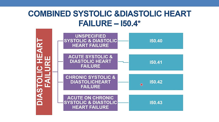


Copyright © 2024 ihoctot Inc.